 |
|
|
SFM
Images of Mildly Trypsinized Chicken Erythrocyte Chromatin Fibers
|
|
|
We have used mild trypsin hydrolysis to analyze the contributions of the linker histone domains and the N-termini of core histone H3 to chromatin fiber structure. Trypsin cleaves the tails of the linker histones and the N-tail of core histone H3 leaving the other core histones intact. Leuba, S. H., Bustamante, C., Zlatanova, J., and van Holde, K. Contributions of linker histones and histone H3 to chromatin structure: scanning force microscopy studies on trypsinized fibers. Biophys. J. 74(6) 2823-2829 1998a PDF file
|
|
| Click on Thumbnail to View Larger Image | ||
|---|---|---|
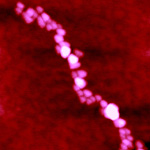 Control Chromatin Fiber | 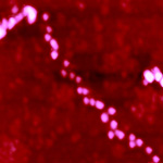 16 min. Trypsinized Chromatin Fiber | 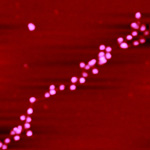 32 Min. Trypsinized |
| 136 KB | 94 KB | 96 KB |
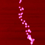 1 Hour Trypsinized | 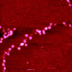 8 Hours Trypsinized | 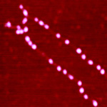 Linker Histone-stripped Control |
| 83 KB | 195 KB | 105 KB |