Physical Molecular Biology Section
University of Pittsburgh Laboratory of Physical Molecular Biology; Sanford Leuba, Ph.D.
researchpeoplegallerypublicationsLeuba lab home
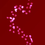
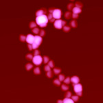
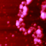
Simulated and Experimental SFM images of Native Chromatin Fibers.
All images are 250 x 250 nm in size.
   
University of Pittsburgh Home University of Pittsburgh School of Medicine Pitt SOM Department of Cell Biology and Physiology Read a bio of Sanford Leuba, Ph.D.
 
Click on Thumbnail to View Larger Image
77 KB73 KB92 KB
A. Computer-generated Image
of a Model Chromatin Fiber.
B. Simulated SFM Image
of the Model Chromatin Fiber in 'A'.
Experimental SFM Image of a
Glutaraldehyde-fixed Chromatin Fiber

Using the following constraints:
(1) the linker histones H1 and H5 help maintain the DNA entry/exit angle on the nucleosome; (2) the DNA linkers between nucleosomes are rigid and straight under the low ionic conditions used here; and (3) the DNA linker length varies within biochemically defined limits (constant linkers would generate a regular helical fiber of nucleosomes); we have been able to model at a first approximation the irregular, three-dimensional character of chromatin fibers.