 |
|
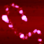
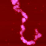
Chromatin Fibers Compact with
Salt |
|
| Click on Thumbnail to View Larger Image | ||
|---|---|---|
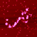 0.1 mM MgCl | 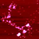 0.1 mM MgCl | 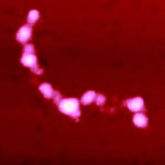 0.2 mM MgCl |
| 93 KB | 90 KB | 82 KB |
 0.2 mM MgCl | 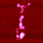 0.3 mM MgCl | 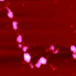 0.3 mM MgCl |
| 81 KB | 80 KB | 83 KB |
 0.4 mM MgCl 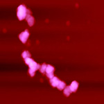 0.4 mM MgCl  0.5 mM MgCl | ||
| 72 KB | 77 KB | 68 KB |
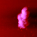 0.5 mM MgCl | MgCl-induced compaction of the extended chromatin fiber, as visualized by SFM imaging. Chromatin erythrocyte fibers, glutaraldehyde-fixed at the respective salt concentrations were dialyzed versus 5 mM triethanolamine-HCl pH 7.0 and deposited on freshly cleaved mica from these solutions. Imaging is in air. Salt concentrations are marked below each panel. Images were taken in collaboration with Dr. G. Yang. | |
| 78 KB | ||