Unfixed Chicken Erythrocyte Chromatin Fibers.
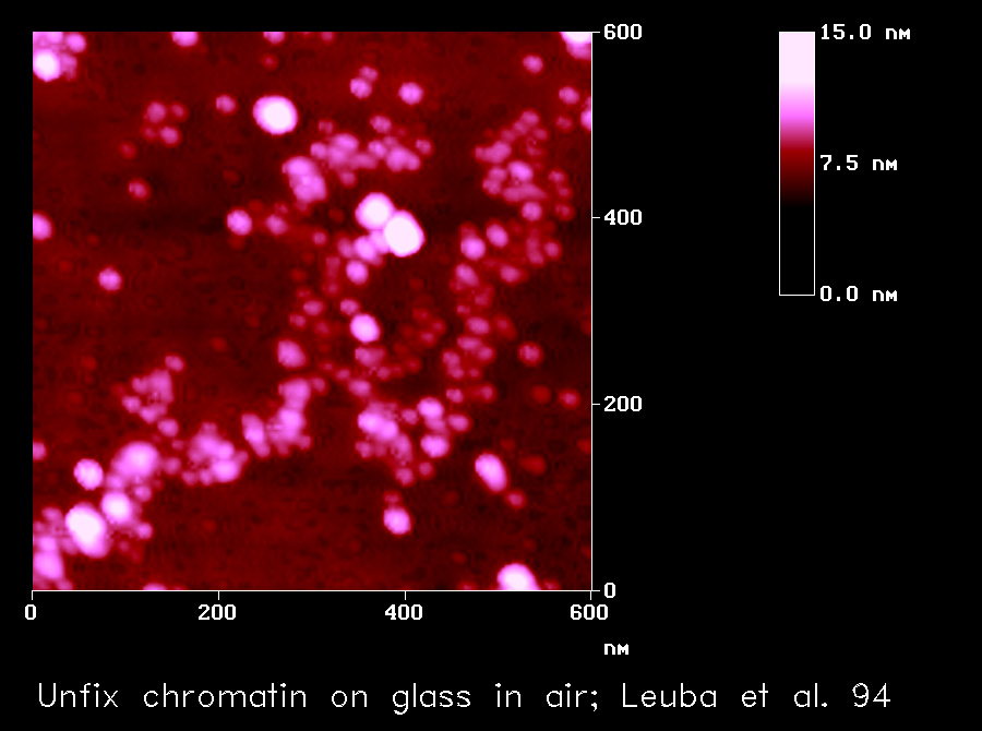
Image size 600 nm x 600 nm.
Image size 600 nm x 600 nm.
Heights are coded by color, with low regions depicted in dark red and higher regions in increasingly lighter tones of red on a height scale from 0 nm to 15 nm.
Leuba, S. H., Yang, G., Robert, C., Samori, B., van Holde, K., Zlatanova, J. and C. Bustamante. Three-Dimensional Structure of Extended Chromatin Fibers as Revealed by Tapping-Mode Scanning Force Microscopy Proc Natl Acad Sci USA 91 11621-11625 1994
| Return to Leuba Home Page |
| Return to Cell Biology and Physiology Home Page |